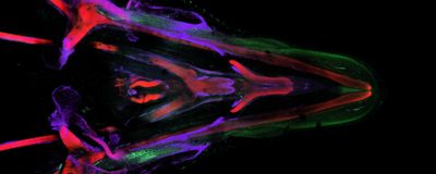
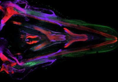
ABOVE: Researchers use chick embryos to investigate how different components of the face and head develop. Evan Brooks
As a kid, Samantha Brugmann, now a developmental biologist at the Cincinnati Children's Hospital Medical Center, had a single Christmas wish: a science kit that came with a microscope and a bee. Using the new scientific tool, she explored the insect’s tiny parts, which sparked an interest in science that inspired her to pursue a career in research. Now in her laboratory, Brugmann and her team peer into microscopes to explore the intricacies of craniofacial development, hoping to understand the molecular and cellular mechanisms in development and disease.
What sparked your interest in craniofacial development?
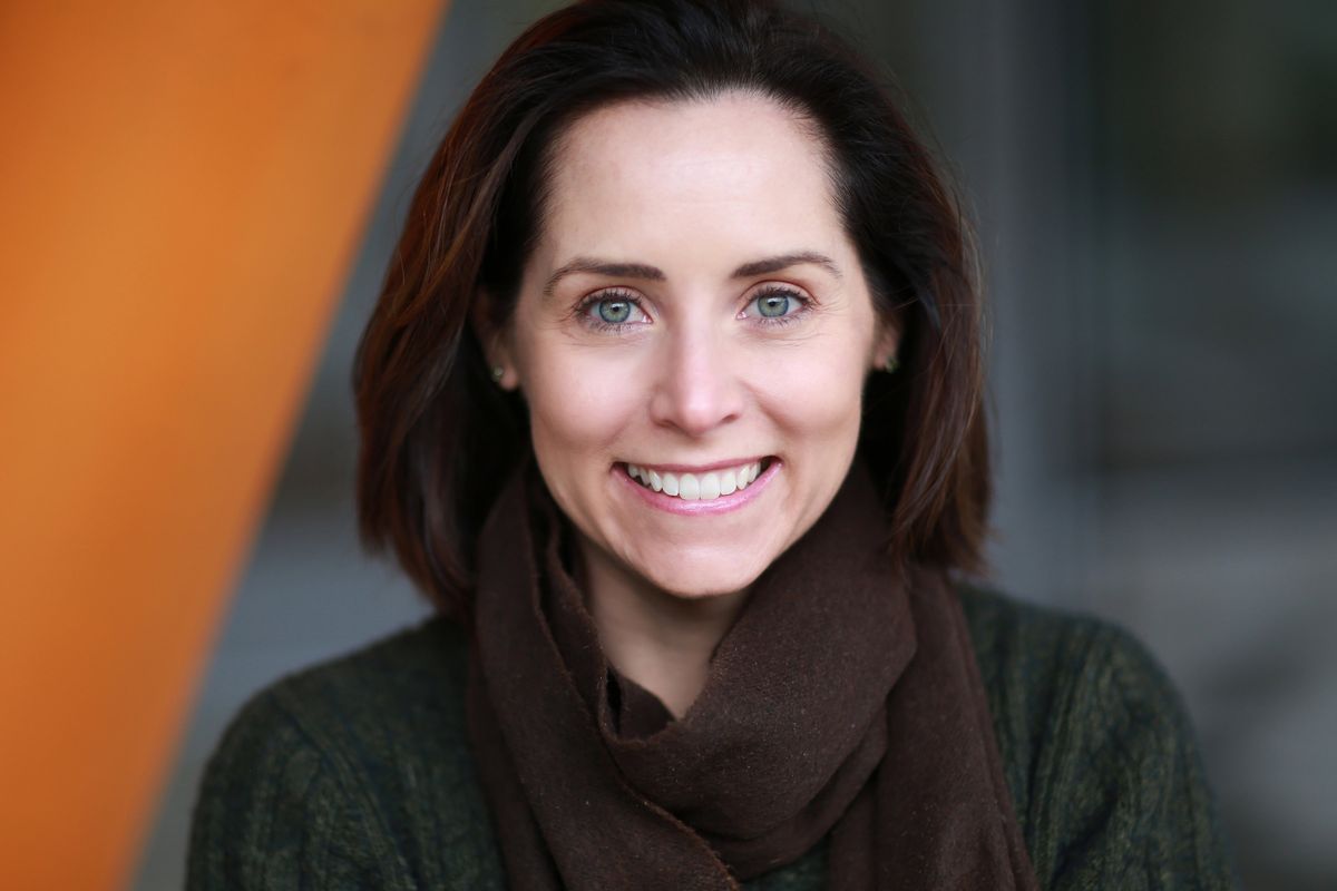
My first contact with the craniofacial field was as a graduate student in the lab of anatomist and neuroscientist Sally Moody at George Washington University. At that time, I investigated the role of certain genes in the development of cranial placodes, which are transient embryonic structures that give rise to all sensory organs.1 As I was preparing to transition to a postdoctoral research position, Sally suggested that I attend a meeting on craniofacial development, where I realized how meaningful this research field is. Unlike other syndromes or injuries that people have, a person with a craniofacial abnormality cannot hide it, and people pass judgments based on our faces. So, if I could help patients with that, it would be impactful for them and their families.
How does your team investigate the mechanisms behind craniofacial development?
We use a few different approaches to study the formation of the head and face, including human induced pluripotent stem cells (iPSC). In this approach, we obtain cells from patients from which we generate iPSC. Then, using specific protocols, we differentiate these iPSC into neural crest cells, which are essential for craniofacial development. We also use mouse and avian embryos to investigate the molecules that go into generating craniofacial structures since this process is highly conserved across different species.
Why are chick embryos an interesting model to study the formation of the head and face?
The chicken model has been seminal for craniofacial development. For instance, neural crest cells were discovered in this model.2 Researchers have also identified naturally occurring variants in chicken flocks that serve as excellent models for human conditions. For example, the talpid avian mutants have craniofacial and limb malformations, and researchers have used them to understand limb and craniofacial development since the 1950s.2
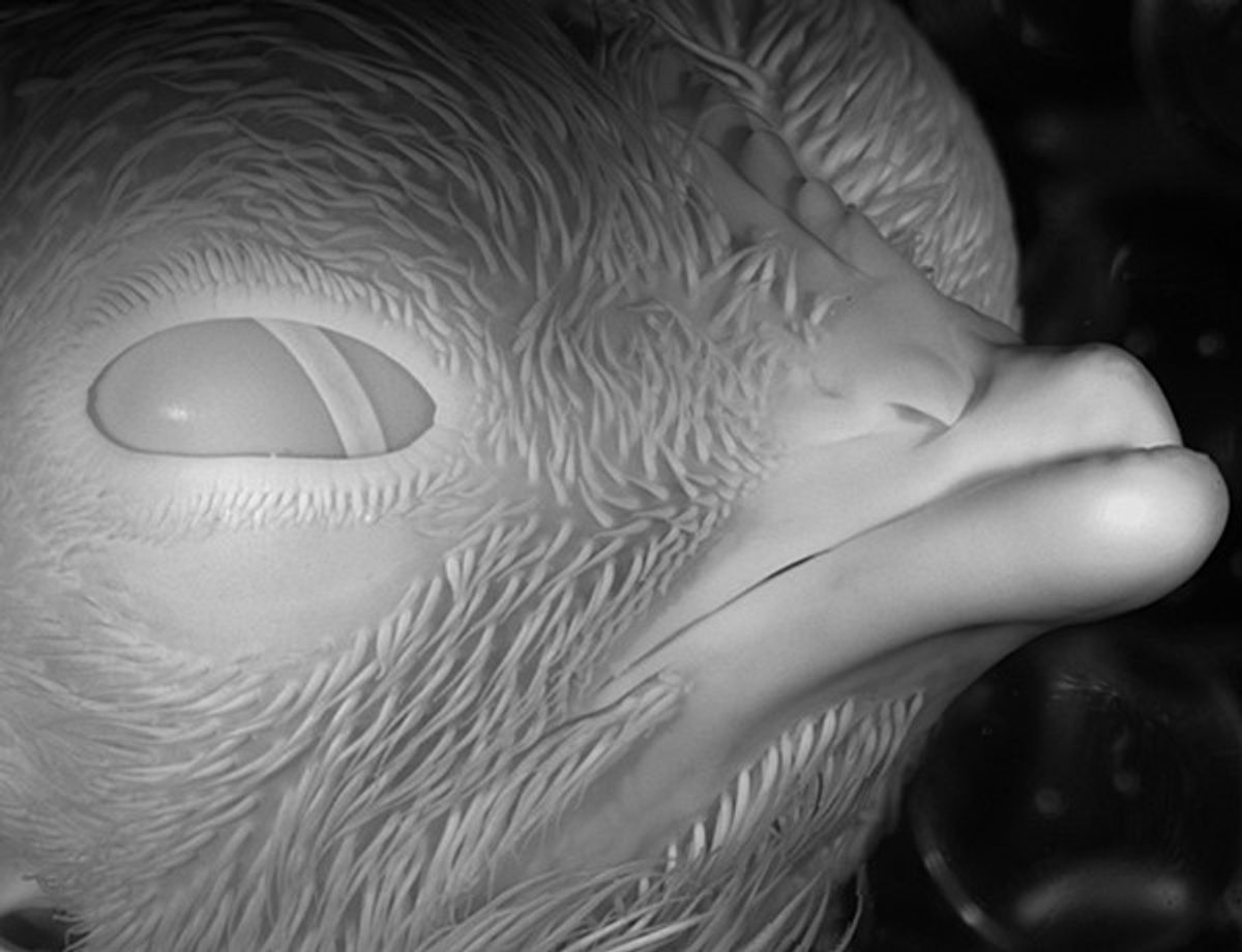
We became interested in one of these mutants, talpid2, based on hints that its craniofacial phenotype was related to primary cilia defects.3 Using whole-genome sequencing in these chick embryos, we pinpointed a mutation in the C2 domain containing 3 centriole elongation regulator (C2CD3) gene, which encodes a protein essential for the primary cilium, revealing a genetic link for talpid2 phenotypes.4 Since mutations in C2CD3 are also seen in patients with oral-facial-digital syndrome, we went on to compare the avian mutant and patient phenotypes and found that they 100 percent phenocopied each other, suggesting that talpid2 is a reliable model for this human ciliopathy.5
What do researchers know about the role of the primary cilia in craniofacial development?
Almost every cell of the human body has a primary cilium. This organelle sticks out of the cell, sensing the environment and transducing that signal. When it comes to ciliopathies, about 30 percent are primarily characterized by craniofacial abnormalities, suggesting that the craniofacial complex is sensitive to the role of the cilium and requires a functional cilium. Although this organelle is essentially described as a molecular signaling hub, we find variation among cilia in different tissues. Recently, we examined this ciliary heterogeneity and found that genes that are important for cilia shape and function are differentially expressed in different tissues.6 This might help explain why we observe a primarily craniofacial phenotype in some ciliopathies and not in others.
What are some of the questions that you plan to explore in the coming years?
We want to keep investigating ciliary heterogeneity, exploring the role of different molecular pathways, particularly those that contribute to bone formation. In the context of craniofacial development, we want to help reconstruct or treat congenital or injured-induced issues, and obtaining skeletal tissue is often one of the biggest challenges. We could apply what we have learned about the primary cilia to the neural crest cells to direct these cells to differentiate into specific cell types, such as bone cells. For patients who need surgical repair, this could mean making bones from the cells that form facial structures, while also preventing any type of rejection since these cells come from the patient’s own body.
What do you enjoy the most about being a researcher?
The excitement of discovering something new is certainly a factor. As a mentor, it is wonderful to see how students evolve from a stage where they need a guiding hand to a stage where they ask questions on their own and take ownership of their projects. Our research also gives us the opportunity to sometimes interact with patients, and that completely changes your perspective on why you do what you do. We can look at chicken eggs all day long and think it's cool, but once you talk to the family and realize how much they want an answer, that’s a level of motivation that makes you work harder to get that data and answers. You want to give something back to them. You want to let them know that somebody is asking these questions to get the answers for them.
This interview has been edited for length and clarity.
Brugmann was nominated for this interview through The Scientist’s Peer Profile Program submissions.
- Brugmann SA, et al. Six1 promotes a placodal fate within the lateral neurogenic ectoderm by functioning as both a transcriptional activator and repressor. Development. 2004;131(23):5871-5881.
- Schock EN, et al. Utilizing the chicken as an animal model for human craniofacial ciliopathies. Dev Biol. 2016;415(2):326-337.
- Brugmann SA, et al. A primary cilia-dependent etiology for midline facial disorders. Hum Mol Genet. 2010;19(8):1577-1592.
- Chang CF, et al. The cellular and molecular etiology of the craniofacial defects in the avian ciliopathic mutant talpid2. Development. 2014;141(15):3003-3012.
- Schock EN, et al. Using the avian mutant talpid2 as a disease model for understanding the oral-facial phenotypes of oral-facial-digital syndrome. Dis Model Mech. 2015;8(8):855-866.
- Elliott KH, et al. Identification of a heterogeneous and dynamic ciliome during embryonic development and cell differentiation. Development. 2023;150(8):dev201237.
