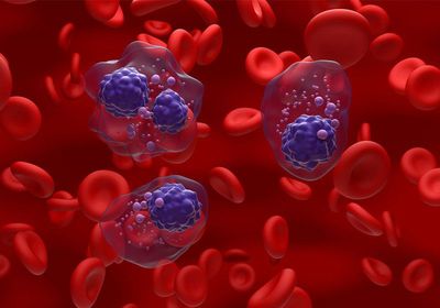
ABOVE: Isolated from patient blood samples, circulating tumor cells are the key to improving myeloma surveillance. ©istock, Nemes Laszlo
Multiple myeloma is a relatively uncommon cancer of plasma cells, where excessive proliferation of these malignant cells within the bone marrow leads to abnormal antibody production. The disease develops from asymptomatic precursor stages, monoclonal gammopathy of undetermined significance (MGUS) and smoldering multiple myeloma (SMM), with a subset of patients progressing from MGUS through SMM to multiple myeloma.1
Irene Ghobrial’s training with Robert Kyle, who she considers the grandfather of myeloma, at the Mayo Clinic sparked her interest in multiple myeloma. She now runs her own laboratory at the Dana-Farber Cancer Institute, where she identifies risk factors that contribute to disease progression and devises innovative approaches for early disease detection. In a recently published Cancer Discovery paper, Ghobrial’s team developed a novel testing procedure called minimally invasive multiple myeloma sequencing or MinimuMM-seq, which allowed them to estimate the patients’ disease burden and detect the emergence of new clones or subclones in a less invasive manner.2
What motivated you to develop MinimuMM-seq?

Many people with MGUS or SMM will never develop multiple myeloma. But every single patient diagnosed with myeloma today must have had these precursor conditions years before. As clinicians, we should be detecting those earlier stages and not waiting for people to have symptoms such as kidney failure, bone fractures, and anemia before treating them. Currently, when we screen asymptomatic patients for the disease and monitor its progression, we perform bone marrow biopsies, which are painful. Because of that, clinicians do not execute the procedure enough times to determine if a patient is responding to therapy. So, although we have amazing drugs to treat patients with myeloma, we are using them completely blind. Additionally, the malignant cells are not evenly distributed throughout the marrow, so the biopsy sample may not be representative of the patient’s condition. After obtaining the samples, we are also still using a very old technology, fluorescence in situ hybridization (FISH), to assess the genomic changes, such as specific mutations or copy number alterations. But FISH fails in many patients, especially in the earlier stages where they do not have enough cells for analysis.
How does MinimuMM-seq work?
In MinimuMM-seq, we examine circulating tumor cells (CTC) derived from patient blood samples. Researchers have shown previously that CTC counts are prognostic, where the more cells that are circulating, the worse the patient’s prognosis. CTC also provide clinicians with information about the malignant cells within the whole body and not just at a single site. We isolated and enriched the CTC from the blood samples using the CellSearch system, extracted their DNA, and performed whole genome sequencing.
What are the advantages of using MinimuMM-seq over bone marrow biopsies and FISH?
Using this method, clinicians do not need to perform a bone marrow biopsy, which is easier on the patients. However, bone marrow biopsies are still likely useful at the beginning to validate the results from MinimuMM-seq. This new technique also allows clinicians to obtain multiple sequential samples to monitor disease progression over time, as well as to examine clonal dynamics including which clones are emerging or disappearing. Additionally, they can acquire information about clones that originate from different areas of the bone marrow rather than from only one area using a bone marrow biopsy. Compared to FISH, MinimuMM-seq displays greater sensitivity and specificity, and it can identify new mutations, translocations, or other genomic alterations that could be critical for understanding multiple myeloma progression and which therapies a patient may benefit from.
What are your next steps?
The current MinimuMM-seq method requires that we use 50 cells or more. We are now working on reducing the number of cells needed to detect minimal residual disease at the very early stages of MGUS. We are also investigating if we can isolate these cells from old frozen samples, as well as comparing the simplicity and sensitivity of this technique to other liquid biopsy methods, such as circulating free DNA. I am hoping in the future MinimuMM-seq will become a clinically validated test, but in the meantime, the method continues to open more doors to a better understanding of multiple myeloma.
This interview has been condensed and edited for clarity.
- Kumar SK, et al. Multiple myeloma. Nat Rev Dis Primer. 2017;3(1):17046.
- Dutta AK, et al. MinimuMM-seq: Genome sequencing of circulating tumor cells for minimally invasive molecular characterization of multiple myeloma pathology. Cancer Discov. 2023;13(2):348-363.


