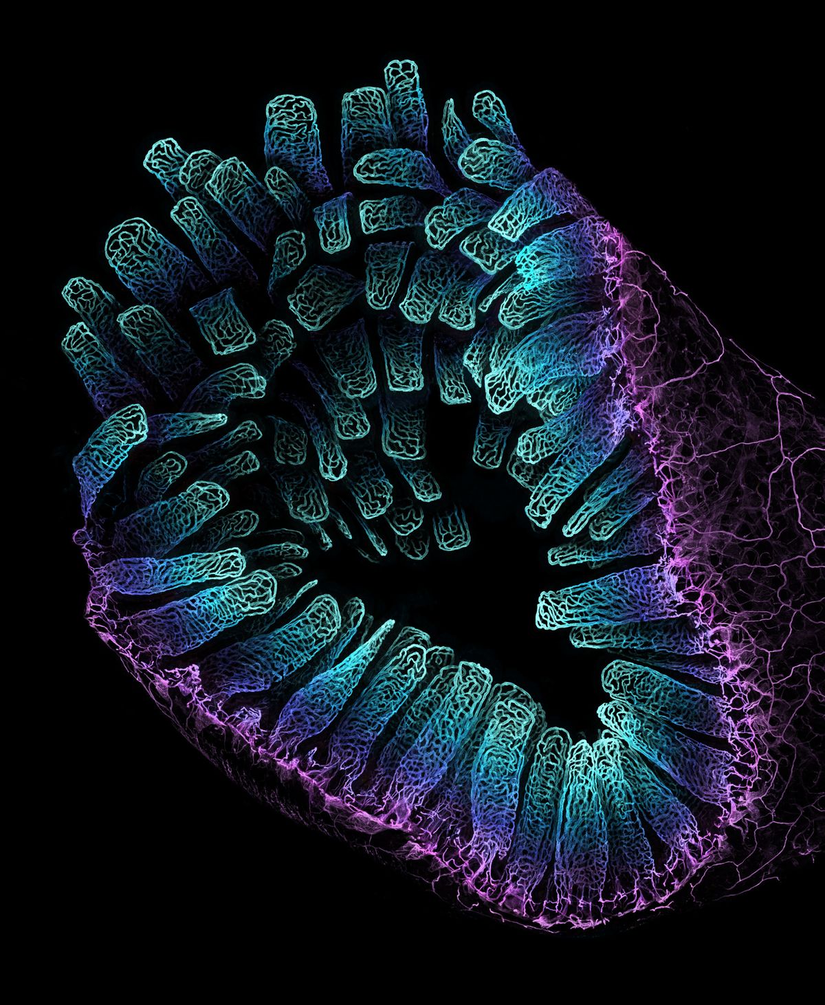
The intestine is like the TARDIS of organs: its surface area is bigger on the inside. Countless villi protrude inward from the intestinal lining, facilitating the absorption of nutrients during the digestive process. Photographer Satu Paavonsalo teamed up with Sinem Karaman, a vascular biologist at the University of Helsinki, to photograph the immense network of blood vessels responsible for this digestive function. Using confocal microscopy, the image features a 10X view of the submucosa (purple) and villi (blue gradient) from mouse intestines. The image took 3rd place at the 2022 Nikon Small World Photomicrography Competition.
